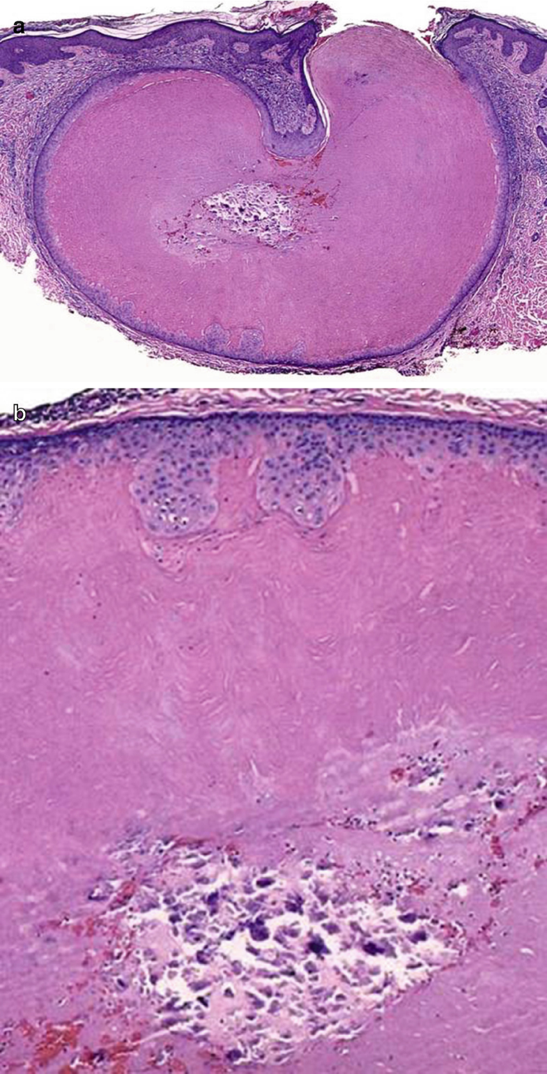
Central Laminated Keratotic Debris

Disease Specific Diagnostics And Medical Management Neupsy Key

Keratosis Obturans Radiology Reference Article Radiopaedia Org

If You Have A Mole At One Of These 7 Places On Your Body This Is What It Means You Will Be Surprised Mole Meaning Natural Medicine Good Interpersonal Skills

Use Once A Week Doubles Hair Growth Combine 1 Tsp Honey 2 Tsp Olive Oil And 1 2 Tsp Coconut Oil W One Half Mashed Avocado Massage Into Dry Hair Wait 10

10 Best Dermatology Images In 2020 Dermatology Wound Care Nursing Medical Knowledge
Nodular And Diffuse Dermatitis Chapter 8 Pearls And Pitfalls In Inflammatory Dermatopathology
A mixed inflammatory infiltrate.
Central laminated keratotic debris.

An Overview Of Hair Follicle Tumours Sciencedirect

Elastosis An Overview Sciencedirect Topics

Eyelid Ento Key

U7esirkrzexpem

What Can Untreated Basal Cell Carcinoma Skin Cancer Do

Skin Chapter 1 Essentials Of Surgical Pediatric Pathology

Kojx0js44ygrym
Skin Springerlink
Http Link Springer Com Content Pdf 10 1007 2f978 3 642 24719 4 16 Pdf
Head And Neck Springerlink
Https Schaberg Faculty Ucdavis Edu Wp Content Uploads Sites 604 2019 07 Skin Tumor 1 Pdf
Https Link Springer Com Content Pdf 10 1007 2f978 3 319 98491 9 Pdf

Tumors Of The Sweat Glands Basicmedical Key
Https Onlinelibrary Wiley Com Doi Pdf 10 1111 J 1600 0560 1994 Tb00243 X

Disorders Involving The Dermis And Or Subcutis E Self Assessment In Dermatopathology
Https Onlinelibrary Wiley Com Doi Pdf 10 1046 J 1365 2133 2000 03382 X
Https Link Springer Com Content Pdf 10 1007 2f978 3 030 10623 2 Pdf

Use Once A Week Doubles Hair Growth Combine 1 Tsp Honey 2 Tsp Olive Oil And 1 2 Tsp Coconut Oil W One Half Mashed Avocado Massage Into Dry Hair Wait 10
Https Encrypted Tbn0 Gstatic Com Images Q Tbn 3aand9gcsylwbcv8p F Gxecsmqz Xekejkeo916ec7kfo5gxmgxtc3p8b Usqp Cau

Epidermoid Cyst Wikipedia
Https Onlinelibrary Wiley Com Doi Pdf 10 1002 Sici 1097 0339 199602 14 1 3c75 Aid Dc16 3e3 0 Co 2 A

Epithelial And Melanocytic Tumors Of The Skin Veterian Key

Home Remedies For Keratosis Pilaris 1 Baking Soda Baking Soda Is An Excellent Homemade Exfoliant This Will Help To Remove The Dead Skin Cells On The Surface
Acitretin In Dermatology Topic Of Research Paper In Clinical Medicine Download Scholarly Article Pdf And Read For Free On Cyberleninka Open Science Hub
Source : pinterest.com
